Physicists Shine Some Light on the
Brain Biologists
and physicists are seeing the mind as never before. Amber
Dance explores how sliced-up brains and X-rays
could help scientists understand diseases like Parkinsons. Illustrated
by Laura Randall and Heather
Gordon. 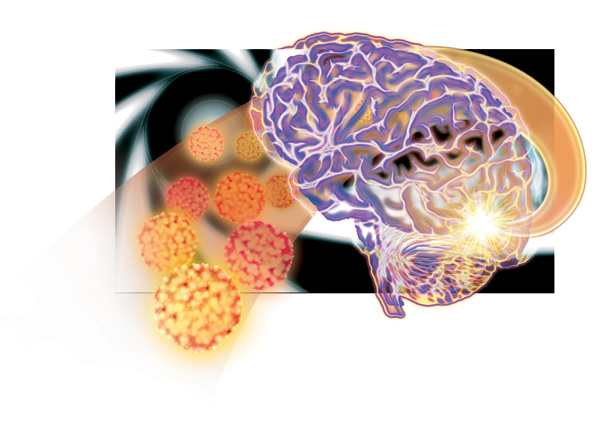
Illustration: Laura Randall Uwe Bergmann explains his take on science with
an old joke: A guy comes home
one night, really drunk, only to discover hes lost his house key.
He looks for it in the small circle of light surrounding a lamppost.
A passerby asks him, Do you really think you left your key there?
I dont know, the drunk replies, but its the only place with light.
For Bergmann, an X-ray specialist
at the Stanford Linear Accelerator Center (SLAC) in Menlo
Park, California, science is like that. We often look in places
where we have some light, he says. Bergmanns
light is a hair-thin beam of sizzling X-rays, shed by a 75-meter-across
circular tube called a synchrotron. In recent years, hes become SLACs
resident guru for anyone who wants to put something unusual in the
beam linefrom ancient manuscripts to body parts to fossils. For
his next trick, Bergmann is focusing his rays on preserved brain
tissue, sliced like a loaf of bread. The
brain research is a collaboration with Helen Nichol, a cell
biologist at the University of Saskatchewan in Canada. Together,
they have put together detailed maps of the metals in a healthy and
diseased brain. Bergmanns beam shed light on Nichol's field of
study: how metals contribute to diseases like Alzheimers and
Parkinsons. For Nichol, the research has
a personal element. Before graduate school, she cared for her
father, who had Parkinsons-like symptoms, and two aunts with
Alzheimers disease. Her inheritance from those relatives allowed
her to pursue her doctorate in her 40s. Now a professor, she studies
how metals, like iron and copper, can build up in a patients snarled brain tissue like a clog in a drain.
Its important to Nichol that she makes a contribution to
medical research. But, she says, science also is a license to
play. With Bergmann, she gets to play with
some fancy toys. Their secret is speedBergmanns X-rays can scan a
brain slice one thousand times faster than other scanning technology.
Armed with maps of different diseases, scientists will now know
where to look for the problems that metals can cause. I dont know
how the heck you would find that by any other means, Nichol says.
Mental metal While
scientists have known for decades that metals are associated with
disorders like Alzheimers, they still dont know whether the metals
cause the disease or result from it. A
healthy brain needs metals. Iron, in particular, is necessary for
neurons and their communication. The brain stores iron in a large
protein molecule: a soccer-ball-shaped cage called ferritin. As
iron atoms follow channels to the center of the cage, they get
oxidizedessentially, they rust. A single ferritin can hold 4,500
iron atoms. It sequesters extra iron, out of the way in the rusted
form, so it cant interfere with the brains chemical reactions.
One section of the brain that uses lots of iron
is the substantia nigrablack stuff, named for its
dark pigment. It sits at the base of the brain, between the ears
at the center of motion control. The substantia nigra
uses iron to produce dopamine, the pleasure hormone that rewards
the brain for activities like eating and sex. Dopamine also helps
to control movement. In people with Parkinsons, dopamine-making
cells bloat and die, and patients struggle to control their bodies.
In Parkinsons and similar diseases, the brain
seems to lose control of iron. Ferritin cant hold it all, and iron
spills out into brain. Unchecked, it starts oxidizing everything
in its path. If you crack an egg and the white part of the egg
starts to solidify, thats an oxidation reaction, says James Connor, a neuroscientist
at the Hershey Medical Center in Pennsylvania. Think of that egg
white as a membrane in your cell. As the membranes stiffen, the
neuron cant function and it dies. Reactive iron can also form
dreaded free radicals, which damage DNA and interfere with other
chemical reactions. In Alzheimers, excess
iron is often mixed up with the plaquesbig globs of unorganized
proteincharacteristic of the disease. In this case, the misplaced
iron may pull other iron atoms away from the neurons that need them,
Nichol speculates. You end up with brain cells that are almost
starving for metal, she says. Until now,
scientists have only been able to study small pieces of brain. They
use chemical stains to color metals in ultra-thin slices, but that
labels only one kind of metal at a time. Or, they dissolve small
chunks in acid or grind them up. They dont have a whole brain map
of metals from any single patient, just bits and pieces from different
brains. Jon
Dobson, a biophysicist at Keele University in the United Kingdom,
uses X-rays to analyze fingernail-sized bits of brain. He hopes
to find brain areas that collect extra iron when diseased, and to
use the information to diagnose Alzheimers in its early stages.
Some of the excess iron forms magnetite; he figures it should be
possible to see it in living patients with magnetic resonance
imaging. But looking at the brain a centimeter
at a time is like trying to appreciate the Mona Lisa with a penlight.
Dobsons group might take all day to run a single sample at Argonne
National Laboratory in Illinois or the Diamond Light Source in South
Oxfordshire. Time on the synchrotron is expensive and hard to get,
Dobson says, so speed is valuable in this kind of research. Better, faster, stronger Enter Bergmann and his team of physicists and
engineers. Theyre all about speed. For a project to reveal ancient
text written by Archimedes (see sidebar), SLAC researchers developed
the hardware to scan faster with X-rays. Instead of taking a
picture, moving the beam, and repeating those steps, they can now
scan smoothly along the sample. Its like graduating from a still
camera to video. They also wrote software to track the beams
position and process the rapid-fire stream of data. Bergmann, 44, is a lanky, athletic German who
strides across SLACs campus at the pace of a speed walker. Hes
also a self-described synchrotron junkie. A senior scientist at
SLAC, he specializes in developing X-ray techniques to study matter
at the atomic scale. His primary research is on the water-splitting
center in photosynthesis, which ultimately powers life on this
planet. With that work and his collaborations, he keeps the X-rays
zipping. I do compete, sometimes, for my own beam time with myself,
he says. His beloved synchrotron houses a
river of electrons, circling at nearly the speed of light. Traveling
electrons spit out X-rays as the beam bends. The tube, about an
inch thick, is surrounded by a set of warehouse-like buildings
crammed with scientific equipment. At 25 stations around the
synchrotron ring, scientists tap those X-rays for their experiments.
The X-rays from the synchrotron bounce through a set of mirrors to
be focused on the sample. Bergmann aims his
X-rays with high precision. He forces them through a metal straw
with a 50-micron holeabout the width of a fine human hair. Its too
thin to see lamplight through the tube. It always takes some time
to fiddle the beam through that thing, Bergmann says. Once aimed, the X-rays hit Bergmanns sample. The
samplebe it brain, parchment, or otherwisesits in a frame that can
move up-and-down and side-to-side, so the physicists move the sample
through the beam as they scan. A regular medical X-ray scan wouldnt
work because there is so little metal to image, so the scientists
have to get fancy. When an X-ray hits a bit of metal, it sparks
the metal to spit out an X-ray of its own. Then, they collect those
secondary X-rays. The
detector is one of the real pieces of magic we have here, says
Martin George, a SLAC programmer who wrote the code to make their
baby run. When the secondary X-rays hit the germanium atoms in the
detector, they produce a spurt of electrons. The computer translates
the electrical signals into an image. With this setup, the team
can take pictures of anything thats got metal atoms in it. With the new equipment, Bergmann first trained his
beam on the Archimedes Palimpsest.
Nichol was using X-rays to scan fly brains at that point. When she
heard about Bergmanns setup, she wanted in. I thought, well, heck,
if he can do a whole sheet of paper, the brain is smaller than a
sheet of paper, she recalls. Theyve got brains
at SLAC Nichol asked a pathologist for
slices of preserved brain from people whod donated their bodies to
science, one Parkinsons and one healthy sample. Preparing the
brains for imaging was easythey simply sandwiched the squishy tissue
between two plastic sheet protectors, like you might find at any
office supply store. The tricky part, Nichol says, was finding a
brand that didnt include any metal in the plastic. Only Itoya brand
sheet protectors would do. Then, Nichol FedExed the brains to SLAC.
Pretty gross, was Bergmanns reaction to the packages contents: a
pale pink slice about an inch thick, with its lobes splayed out
like petals on a flower. Although SLAC
houses cutting-edge research from every kind of science, Brain Day
was an exciting event at the synchrotron lab known as SSRL. George
remembers that Saturday: After dropping his wife off at the airport,
he just had to stop by the lab. Hey, theres slices of brain at
SSRLIve got to take a look, he recalls. At
SSRL, one can stroll the catwalk and look down into what one of
Bergmanns collaborators described, quite accurately, as an electronics
junkyard. Wires and foot-thick pipes snake across the room.
Cobbled-together devices sprout dozens of plugs, and power strips
line up in ranks of six or more. Racks of electronics gear lurk
around every corner, blinking their many lights. Much of the
equipment hides in a snug coat of aluminum foil, as if trying to
ward off alien brain waves. (Actually, the foil insulates the gear
and protects it from dust.) The occasional physicist stares into a
computer terminal or laptop. At each station,
the X-rays shoot off the synchrotron into a hutch, a sort of walk-in
closet holding a table for the instruments. Closing the equipment
in the lead-lined hutch protects the scientists from the X-rays,
which can cause cancer in high doses. When
its time to fire up the detector, Bergmann and his colleagues haul
their equipment from the corner where it squats, covered in a plastic
sheet, to the hutch. Carefully, and with the help of some duct
tape, they set up the gear amid a riot of brightly colored wires.
Bergmanns detector is a million-dollar piece
of equipment that looks like something out of a low-budget sci-fi
flick. Its a tube a little wider than the brain itself, covered
in foil. Even though it may look like its been put together with
tape and stringit hasthat doesnt necessarily mean weve been careless
with what we do, George says. In fact,
Bergmann is a perfectionist who refuses to even look at an image
until hes checked all the equipment. I really get my hands dirty,
he says. With everything just right, they
close the hutch door and start the scan. A few hours later theyve
got a map, not just of iron, but also of a host of other metals.
The X-ray beam hits each spot for a few milliseconds, crisscrossing
the sample so fast that it isnt even damaged. Nichol can return
the brain to the pathologist unharmedan important factor if she
wants to image rare samples. As expected,
the substantia nigra of the Parkinsons patient was black
indeed, clogged with iron. Nichol also saw halos of iron surrounding
the blood vessels in the diseased tissue. Scientists had suspected
blood vessels were involved in disease, but they had never seen
this evidence before. In future studies, Nichol hopes to identify
which form of iron surrounds the blood vesselswhether its reactive
or rusted. To Nichol, most surprising was
the relationship between iron and zinc. In both brains, areas high
in iron were low in zinc, and vice versa. Some zinc-containing
proteins bind to dangerous metals and protect against oxidative
damage, so scientists think they might block the symptoms of
Parkinsons disease. Nichol intends to follow up on this finding
as well. A brain a day Those first pictures proved to the scientists
satisfaction that the technique works. SSRL administrators also
were pleased. Theyre very keen on this, Nichol says, because of
the health implications. She expects to get more beam time, and
she has permission to look at ten more samples. Nichol is starting by imaging brains with a variety
of diseases. The most exciting discovery, she says, would be to
find a role for metals in a disease for which no one has yet
considered them. In that case, she says shed need about a dozen
brains to convince other scientists. With equipment that scans a
brain every six hours, that is now something she can envision doing.
And the technique is not limited to brainsany metal-containing organ
could eventually get its turn in front of the X-rays. Another possible application is animal research.
Scientists trying out new drugs, for example, could use the speedy
scans to look for neurological effects. In just two days, Nichol
estimates, they could get through 25 mouse brains. At the time of writing, Nichol had not yet shared
her results with other biologists. However, scientists have wished
for this kind of data. Complete maps of individual patients are
not available, James Connor and co-authors wrote in a 2004 article.
Moreover, data on iron in some areas that are seriously impaired
in Alzheimers disease and Parkinsons disease are still missing.
With Nichols brains and Bergmanns machine, those maps may come out
soon. Nichol expects the new maps will
help scientists understand neurological diseases, and perhaps reveal
ways to slow or halt the damage metals can do in the brain. One
possibility is chelation therapy. Chelators are molecules that
act like sponges for metal atoms, sopping them up and keeping them
from causing trouble. The molecules cage metal ions with more than
one chemical bond. The kidneys and liver then remove the chelator-metal
pairings from the bloodstream. Chelation treatments are used for
heavy-metal poisoning and Wilsons disease, in which a persons body
has too much copper. In the 1980s, scientists
tried chelation therapy for Alzheimers disease, but the project
fizzled. The treatment required painful intramuscular injections,
says Mark Smith,
a neuroscientist at Case Western Reserve University in Cleveland.
Ive heard, anecdotally, that patients who have thalassemia
[a disorder including iron overload] would prefer to die of thalassemia
than have the treatment, he says. Chelation
therapy for neurodegenerative disease may get a second chance.
Pills have replaced the painful shots, and an Australia-based
company, Prana, is doing clinical trials. No chelation treatments
have yet made it to the pharmacy shelves. The problem so far has
been that the chelators are too good, Connor says. They dont
differentiate good and bad iron. Any potential
treatment coming out of Nichols research is years away. We are
just telling them where to look, Bergmann says. For scientists who
used to squint at tiny parts of the brain, suddenly the floodlights
are on, and theres so much to see. Top
Sidebar: 21st
Century Look at a B.C. Book 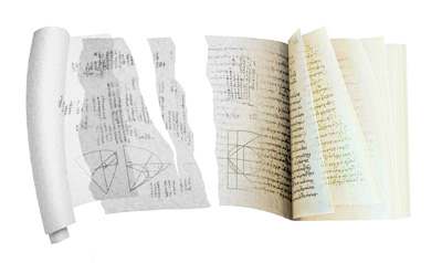 | Illustration: Heather Gordon |
The monk Johannes Myronas needed
parchment. He wished to construct a prayer book. But in 1229, parchment was expensive and hard to
get. So Myronas recycled an old scroll, scraping off the dark iron
gall ink and writing his prayers on top. The practice was called
palimpsesting, from the Greek for scraped again. And in that moment,
musings by the famous mathematician Archimedes of Syracuse, who
lived in the third century BC, were nearly lost forever. Eight centuries after Myronas obliterated them,
it took a modern scientist to unveil those thoughts. Uwe Bergmann,
an X-ray specialist at the Stanford Linear Accelerator Center, used
his ultra-fast scanner to map traces of iron from the erased ink.
The book had traveled from the monastery, through
the hands of forgers who damaged it, to Christies auction house in
1998. An anonymous buyer offered the book to scholars, who partnered
with Bergmann. You can imagine that if you tell some owner of a
two-million-dollar book that we will put this book into a beam a
million times brighter than the sun, he will be very delighted and
give it to you right away, Bergmann says. And in fact, he was.
Using the synchrotrons powerful rays, Bergmanns
technique lit up the iron atoms in the ink. This allowed scholars
to read the original writings: at least seven treatises by Archimedes
and work by others. Of these, two Archimedes works had survived
nowhere else. The palimpsest contained the only known text of the
"Stomachion," in which Archimedes describes
his favorite puzzle game. The object is to use different-shaped
tiles to build other shapes, such as people or animals. In addition, Bergmanns beam illuminated the only
copy of Archimedes letter to his friend Eratosthenes, the librarian
in Alexandria. In it he describes his Method of Mechanical Theorems,
in which he used mechanical metaphors to do mathematical calculations.
The Archimedes project brought Bergmanns X-ray
scanner to the attention of other scientists. In addition to his
work on brains, he plans to scan a fossil
Archaeopteryx, the feathered dinosaur that bridges the divide
between reptiles and birds. He and his collaborators hope to learn
about the animals skin by studying the elements it left behind.
Top
Biographies  Amber Dance Amber Dance
Sc.B. (biology
with honors) Brown University
Ph.D. (biology) University of
California, San Diego
Internship: Nature (Washington,
DC)
The FBI tour guide stopped in
front of a window to a molecular biology lab and asked whether
anyone knew what the letters DNA stood for. The adults shook their
heads, but a small voice from a 12-year-old piped up: De-oxy-ribo-nucleic
acid. That 12-year-old was me, a science nerd from the beginning.
I also devoured any writing in front of mewhether magazines, adult
novels my mom never should have let me read,
or the backs of cereal boxes. I
followed my love of science to a biology degree and a Ph.D. But
somewhere in the middle of an endless search for mutant bacteria,
that love withered. Im thrilled to trade my pipette for a pen, and
I hope to recapture my fondness for science while letting someone
else do the tedious mutant hunts. . . .
. . . . . . . . . . . . . . . . . . . . . .
. . . . . . . . . . . . . . . . . . . . . .
. . . .  Laura
Randall Laura
Randall
B.S. (ecology and systematic
biology) California Polytechnic University, San Luis Obispo
My concentration in wildlife biology at Cal
Poly, my research experience, and my job experience as a veterinary
assistant further cultivated my love and understanding of the natural
sciences. After 9 months of intense instruction, artistic growth,
and lots of fun in the Science Illustration Program, I am now living
in San Diego to pursue an internship in the entomology department
of the San Diego Natural History Museum. . . . . . .
. . . . . . . . . . . . . . . . . . . . . .
. . . . . . . . . . . . . . . . . . . . . .
.  Heather
Gordon Heather
Gordon
BA, Literature/Creative Writing,
University of California, San Diego
But
it is of course easier, when we have previously acquired, by the
method, some knowledge of the questions, to supply the proof than
it is to find it without any previous knowledge. Archimedes (to Eratosthenes), from
The Method Maybe the same could
be said about the relationship between scientific and aesthetic
discovery. Anyway, it has been my pleasure to consider. And to
draw. Enjoy! Top
|