 |
|
| |

| Impelled by the tragic plight of paralyzed victims of spinal-cord injuries, scientists move ever closer to unlocking the mysteries of nerve development and regeneration. |
|
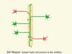
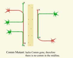
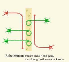
It’s the eighteenth day of conception. In the darkness of the mother’s womb, a groove begins to form in the embryo. The groove deepens and its lips rise and curl toward each other. Clumps of cells growing on the lips escape just before the groove closes into a tube. They crawl away and take their positions, like sentinels in the developing body. Soon, these cells and those inside the tube reach out for others with snake-like extensions to touch, connect, and wire an exquisite network – a trillion cells, each conversing with a thousand others. These cells are nerve cells, or neurons. The tube is the budding brain and spinal cord, and their gloriously fragile network is our nervous system, the most complex of systems in the human body. The wiring starts when isolated neurons hesitantly extend their tentacle-like protrusions called axons. Lone, eager axons sniff their way to their targets, stretching toward or veering away from chemical cues around them. These pioneering axons lay themselves down like tracks. Those that start later ride along, using the tracks when necessary and abandoning them when not. Some axons miss their targets; their fate is death. After decades of research, scientists now are uncovering some of the processes that accurately guide these axons through the embryo. Using genetic experiments and surgical scalpels, researchers have hunted for and found molecules that pull, push, or hem in axons; genes that regulate axon movement; and clockwork mechanisms that require precise timing on the part of growing neurons. They have discovered that creatures as varied as fruit flies, roundworms, and rats have strikingly similar mechanisms, suggesting a common basis for nerve growth in all species. Scientists hope that learning precisely how nerves grow in lower animals may one day help them regenerate damaged nerves in humans. Neurotic Neurons Scientists weren’t always this optimistic. In fact they were divided on the basic question of just how the nervous system was wired. In the early 1960s scientists thought that axons grew and reached targets randomly. They believed the nervous system somehow checked itself after the wiring was complete, keeping valid connections, and pruning erroneous ones. In this scenario, an axon extending from a neuron would meander and find some target, say a muscle cell. After the connection had been verified, the neuron would have to function as one that controls muscles. This situation is not unlike picking up the telephone, dialing randomly and reaching Bob’s father instead of your own. Now imagine being forced to be Bob because you’re talking to his father. Because that’s what a neuron would have to do: permanently adopt an identity depending on the target it reached. "The theory didn’t make sense," says Corey Goodman, a neurobiologist at the University of California, Berkeley. It didn’t make sense to Roger Sperry either. In 1963, Sperry, a neurologist at the California Institute of Technology in Pasadena, proposed the idea that specific molecules chemically attract axons, pulling and guiding them to their targets. Sperry did not claim to know what the molecules were, just that they had to be there. However, colleagues slammed Sperry for his ideas. Even as recently as 1985, a group of eminent scientists collectively argued against his theory. As for the mysterious attractive molecules, explains Goodman, the scientists said, "Show them to us. Where are they?" Soon thereafter, Goodman and other researchers began to validate Sperry’s idea. Their studies found that, when confronted by groups of cells on its path, the tip of the axon, known as the growth cone, would touch them all but would be attracted only to a certain group. When researchers surgically removed the cells that attracted the growth cone, they found that it did not move toward the remaining cells; it was very particular in its taste. By the mid-1980s, researchers were convinced that growth cones were making very specific decisions about how they moved from the source neuron to the target cell, and that specific molecules were guiding the decisions. Meanwhile, researchers were discovering that axons navigated their paths one small segment at a time, much like commuters going from their homes to their offices travel on different roads and freeways along the way. At the end of each segment, the growth cones on axons had to choose which way to go next. And, at the same intersections, different growth cones made different choices. "Why do some cells turn left and other cells turn right? If we actually understood that at a deep level for a number of different kinds of decisions, we would understand a lot about brain wiring," says Goodman. Midline Crisis: To cross or not to cross To study these decisions, researchers focused on a special row of cells in the embryo called the midline. These cells lie, moatlike, in the middle of the developing nervous system, dividing it up into mirror-symmetric right and left halves. As it turns out, almost every neuron that wires the brain and the spinal cord has to confront the midline and make decisions at some point in its lifetime, says Goodman. As axons jostle with the midline, some stay on their half of the embryo, while others cross over to find targets on the opposite side. The axons that cross the midline are called commissural axons (from the Latin word commissura, which means "a joining"). The midline became the target of a fortuitous collaboration between researchers in Goodman’s lab and the lab of cell biologist Jonathan Rothberg at Yale University. Their genetic experiments with fruit flies, in which they bred mutant versions by tinkering with one gene at a time, revealed something spectacular. In one particular mutant, which lacked a yet-to-be-named protein, the axons from either side of the nervous system converged on the midline but never crossed over. Instead they ran up and down the center, slitting the organism, as it were. The missing protein responsible for this miswiring was named Slit, and the fruit fly strain was called the Slit mutant. That was in 1990. Neurobiologists would have to wait nearly a decade before someone could explain why axons collapsed onto the midline in the absence of Slit. Next, Goodman and his team began an exhaustive search for genes that affect midline crossing in fruit flies. In 1993 they found two. Lacking the first gene, the flies had no commissures, or axons crossing the midline. They named the gene Commissureless, or Comm. Without the second gene, the axons crossed the midline but came right back and helplessly circled around in it. To Guy Tear, an English researcher in Goodman’s lab, they looked as if they had been caught in the British traffic circle: the roundabout. The gene was called Roundabout, or Robo. Meanwhile, across the bay from Berkeley, neurobiologist Marc Tessier-Lavigne and his team at the University of California, San Francisco, were studying axons crossing the midline in rats. In a petri dish experiment with axons and midline cells, the axons would turn from a distance and head toward the cells. Something in the midline cells was attracting the axons. Eventually, in 1996, the researchers purified and identified the substance – a protein. Tito Serafini, a postdoctoral fellow in the lab, searched for a name that suggested the idea of guiding. He settled on the Sanskrit word "netr," for "one who guides," and named the protein Netrin. The stage was set for uncovering more complex mechanisms that guided axons to their targets. In 1998, Thomas Kidd, a postdoctoral fellow in Goodman’s lab, was indulging in one of his favorite activities – dissecting fruit flies to peek at their nervous systems. He discovered that axons crossing the midline had no Robo proteins, which were produced by the Robo gene, but axons growing along the midline did. "It was a tremendous moment in the lab, seeing Robo on two lines (of axons) on either side of the midline, and not on any of the axons crossing," says Kidd. It was clear to him that axons with Robo on their surface kept away from the midline: Something in the midline was repelling the axons. Kidd surmised and eventually proved that Comm proteins in the midline suppressed Robo on some axons, allowing them to cross over. The next experiment hit the nail on the head. Kidd was studying genetically engineered, or mutant, fruit flies with abnormally high levels of Comm proteins. To his surprise, he found that the excess Comm destroyed almost all the Robo proteins on the axons. Whatever repelled the axons by acting on Robo had no effect now. Axons that normally stayed away from the midline now came crashing in, and went up and down the length of the flies, never leaving the center. "This was an absolutely crucial observation, because this (fly) looked like the Slit mutant," says Kidd. In the Slit mutants that Goodman and Rothberg had seen in 1990, the absence of Slit caused axons to collapse onto the midline – exactly what Kidd was observing now. In what he calls his "most exciting moment in science," Kidd had a flash of insight: the Slit protein was the midline repellent that kept some axons away. "It was fantastic," says Kidd. "It’s one of those things where you know you are right. And I kept it a secret for three weeks. Corey (Goodman) was on safari in Africa at that time. I told him when he came back that I knew what Slit did and he believed it straight away." Kidd and fellow researchers then went on to characterize how Comm, Robo, and Slit proteins, in an elaborately choreographed sequence, worked to either keep axons away from the midline or help them across. Since then, researchers in neurobiologist Mu-ming Poo’s lab at UC San Diego and Tessier-Lavigne’s lab at UC San Francisco have found that Netrin in the midline could both attract and repel axons. This depended on the kind of proteins that grew on the growth cone’s surface. A basic principle of axon guidance had emerged: that the same molecule, Netrin in this case, could both attract and repel. As researchers discovered more molecules, a more coherent picture was forming. Apart from the two types of midline proteins – long-range attractants like Netrin and long-range repellents like Slit – they found other classes of molecules operating elsewhere in the nervous system. Proteins were implicated in short-range repulsion; a repulsion so drastic that at times it caused the growth cones to shrink. Other large families of molecules controlled short-range attraction. But a unity is emerging at the molecular level, says Tessier-Lavigne: attractants can repel, and vice versa; and the same family of molecules can act from near or afar. After all the agonizing decisions a lone axon makes at the midline, the axons that follow have it easy. Axons that come later tend to physically cling to these "pioneer" axons, like hikers who stick to well-worn paths. Slowly, as more and more axons cling to each other, they becomes bundles of axons. Eventually, as they near their targets, or their destination requires a different path than what their bundle offers, the axons separate from the group – like a hiker who, after days of walking with others, decides to strike out on his own. Researchers have found proteins that cause axons to stick to each other and proteins that cause them to part ways. Clockwork Death As trillions of axons grope around in the developing embryo, what is it that keeps them from making catastrophic misconnections, like hooking up our taste buds to the visual center in the brain? As is often the case in science, serendipity provided an answer. Tessier-Lavigne and former postdoctoral fellow Hao Wang were studying axon growth toward the base of the midline, called the floor plate. One experiment had axons growing toward floor plate cells, another had axons growing toward Netrin proteins extracted from the floor plate cells. Normally, they stopped the experiments after 40 hours of observation. Except once. "I was being lazy that day. I didn’t throw them out. I came back a few days later, and rather than toss them out, I had a look at them, and it was really remarkable," says Tessier-Lavigne. "In the ones with floor plate cells, the axons were healthy, and in the ones with the Netrins, the axons had shriveled up and died." Something in the floor plate cells had kept the axons alive. Whatever this substance was, it wasn’t needed for the first 40 hours of the axon’s life. Further research confirmed that axons, once they reach the floor plate, become dependent on those cells to stay alive. The life-sustaining chemical is yet to be identified. Researchers have known for decades that axons, once they reach their final target, need chemicals from the target to stay alive. What was different about this finding was that the floor plate was not the final target, but an intermediate one; the axons were just passing through. Tessier-Lavigne believes this is a kind of "intermediate control" system. He named it "en passant," French for "in passing." The reasoning goes something like this: In large organisms such as mammals, axons may grow for up to a week before reaching their targets. If they stray early on and miss their intermediate targets but continue growing, they might eventually find some alternate target, creating a miswired neural circuit. The "en passant" mechanism acts as a checkpoint. Axons given to wanderlust miss these checkpoints and die, like a marathon runner who must "report" to every check station along the way, or risk being disqualified. The Unbearable Likeness of Being While researchers have discovered and studied the "en passant" mechanism only in rats, they have found that many of the mechanisms and molecules guiding axons exist in animals as diverse as fruit flies, roundworms, rats, and other vertebrates. Whether long-range or short-range, attractants or repellents, all or some of these molecules exist in many species, including humans. Slit genes exist in fruit flies, roundworms, and vertebrates. The Robo-Slit interaction that Goodman’s team uncovered in the fruit fly also works in vertebrates. This suggests that evolution has preserved mechanisms from an earlier era, improving upon them for increasingly complex organisms. But neurobiologists took a long time accepting that a human being’s nervous system might be wired like that of a fruit fly. Goodman thinks it was because of our pride. "We like to think we are something special," he says. "As far as we know, going back to the first multicellular organisms, all these organisms have nervous systems. And they all evolved from each other. Was there some place where evolution gave up on brains as it had made them already, and suddenly decided it’s going to make nervous systems in an entirely new way? Of course not. That’s rubbish. In terms of genes, we are not that different from a fruit fly or a frog. That’s a little humbling." Researchers are delighted by these findings. It allows them to study simple nervous systems in fruit flies and roundworms and extrapolate their results to humans, knowing that similar mechanisms may be at work. While axon-guidance mechanisms may be similar among species, there is one phenomenal difference between the nervous systems of warm-blooded creatures such as humans and those of cold-blooded animals. A human’s brain and spinal cord, which make up the central nervous system, both have nerves that cannot regenerate once they are severed. But cut the nerves in a frog and they grow right back. The need to regrow human nerves damaged by spinal cord injuries or brain traumas has driven some of the basic research in neurobiology. Research in the early and mid-1980s focused on finding attractive molecules to coax severed nerves to regrow. But that didn’t work. Then in the late-1980s, researchers found that the central nervous system in adult humans is awash in repellent molecules. These molecules hem in axons and prevent them from growing. There may be an evolutionary reason for this, speculates Goodman. "When we were selected by evolution in the Serengeti plain, we couldn’t run faster than other animals," he says. "We didn’t have bigger jaws, bigger hands. But we were cunning. We were selected for our brains. We have this very plastic brain that allows us to change and to learn. But if you just allow that whole brain to continually sprout and change, it’s going to miswire itself. So somehow, evolution had to evolve a set of principles, if you will, where the basic wiring diagram is held fast, and the endings, or fine details, are allowed to sprout and grow." Neurobiologists think that the repellents stabilize the fundamental network of neuronal connections, keeping its complexity intact. The cutting edge of axon-guidance research is now focusing on how to overcome these repulsive forces acting on growth cones. Goodman thinks there is a mechanism inside a growth cone that somehow sums up all the molecular forces acting on it. If the net result is repulsive, it puts on the brakes, stopping the axon. If it’s attractive, it steps on the accelerator, moving the axon forward. The idea, Goodman says, is to find a way to floor the accelerator in spite of the repulsive forces acting on the axons. Mu-ming Poo and Tessier-Lavigne may have floored such an accelerator. While studying frog embryos in the lab, they found a way to repel or attract axons to the same external chemical cues. They did this by varying the level of chemicals called cyclic nucleotides inside the growth cone. In general, lowering the level of these cyclic nucleotides repelled axons, while raising them attracted the axons. Whatever the mechanism inside the growth cones, these chemicals could somehow step on the brakes or the gas. Scientists have yet to confirm these findings in live animals. The "most exciting possibility" raised by this research is that damaged nerves could be regrown by drugs that raise the level of cyclic nucleotides in injured axons, wrote neuroscientist Pico Caroni of the Friedrich Miescher Institute in Basel, Switzerland, in a commentary in Science magazine. Tessier-Lavigne is also very optimistic. "I think it’s realistic to hope that in 10 to 12 years there will be drugs to help people with spinal cord injuries," he says. But he adds an important caveat. While it may be possible to get nerves to regrow, it’s not certain they’ll make the right connections. If they don’t, it might be worse than having no growth. But, says Tessier-Lavigne, "there are lots of indirect reasons to think that axons will be able to reconnect with the right kinds of targets." Then there is the question of the extent of regrowth needed for a patient to recover. "Christopher Reeve is on a respirator. We might get some regrowth that might get him off the respirator. Will we get enough regrowth that so he can move his shoulders? That might still be realistic. Will he able to move his hands or his legs? That’s a tougher call because it requires the axons to grow more and more down the spinal cord to regain those functions," says Tessier-Lavigne. "To get a measure of regrowth is feasible. How much, time will tell." Currently, in the event of a spinal cord injury, doctors give patients massive doses of anti-inflammatory drugs. This has to be done in the first eight hours after the trauma, because soon after that the patient’s own immune system senses the damage, and starts attacking and killing the axons. The drugs to regrow axons may have to be given within this eight-hour window to be effective, Thomas Kidd speculates. Whatever the eventual outcome, neurobiologists have come a long way since Sperry proposed, in 1963, that molecules must be attracting and guiding axons diligently to distant waiting cells. In embryos of rats, worms, flies, and humans, they have discovered that these axons act much the same, harking back to primordial pioneers that wired the first nervous systems. |
 |
||
|
Anatomy of a nerve cell
|
|||
 |
|||
 |
|||
 |
|||
- BIOs
- WRITER Anil Ananthaswamy
- B.Tech (EE), Indian Institute of Technology, Madras, India. MS (EE), University of Washington, Seattle, USA
Internships: Santa Cruz Sentinel, Santa Cruz, California; New Scientist, London, UK
- ILLUSTRATOR Jennifer Johansen
- B.A. Biological Illustration, Iowa State University
Internship: Scientific American, New York