Cultivating Autism
A neurobiologist grows brain cells to explore the roots of autistic behavior. Jane Palmer peers into his lab dish. Illustrated by Eva Langhorst and Jane Kim.
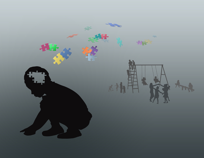
Illustration: Eva Langhorst
When Ricardo Dolmetsch learned his son had autism, he knew his life would change. Little did he know, so would his research.
In his neurobiology lab at Stanford University, Dolmetsch studied how the complex wiring and firing of neurons shaped the human brain. Although he described his work as “an entirely curiosity-driven, intellectual pursuit,” it had served him well. At 37 he’d earned a string of awards, including the prestigious Young Investigator Award from the Society of Neuroscience. A fast-talking firecracker of ambition, the native Colombian was riding the wave of academic success.
Then in 2007, his son’s diagnosis stopped him in his tracks.
“When I started thinking about what was going to happen to this kid in the future, it was the worst feeling in the world,” says Dolmetsch, his speech slow and faltering. “It was such a terrible, powerless feeling.”
Determined to do something, Dolmetsch scoured the literature, learning all he could about the disorder. What he found was disheartening. “I encountered a large amount of behavioral data, a small, but growing amount of genetic data, and almost nothing in between,” he says.
As a scientist who had studied neurological diseases such as schizophrenia, Dolmetsch knew this gaping hole in the research didn’t bode well. Without knowledge of the complex circuits of neurons involved in autism, hopes for a treatment would be slim. The discovery, however, proved life-changing.
“It galvanized me into action,” says Dolmetsch, an athletic man with a passion for soccer and running. “I realized I had spent all my time just following my curiosity, and now maybe I should do something to help somebody else.”
He began to consider how to use his own research to study the disorder. His lab explored how the firing of an electrical impulse from one microscopic neuron to another launches a flood of calcium into the cell, changing its behavior. Coincidentally, scientists had already discovered a connection between the genes that control this cascade of interactions and autism. Dolmetsch’s work and autism were linked.
His main concern was not switching the focus of his lab, but how to overcome one of the biggest obstacles in the field: getting access to the neurons of autistic children. Scientists have made great progress in studying cancer because they can do tumor biopsies. But taking a biopsy from the brain of a living child is unthinkable. If he wanted to understand the origins of autism, Dolmetsch knew he would need to get access to those brain cells. But how?
Autism’s tangled roots
Autism has puzzled scientists for nearly seven decades. In 1943, Leo Kanner of John Hopkins University published a paper [1] describing the behavior of eleven children who seemed to have shown no interest in interacting with other people from birth. These same children were also obsessively interested in particular aspects of their environment. He called the disorder autism, from the Greek word “autos,” meaning “self.” The children he observed were creating an “isolated self.”
Although the definition of the disorder has changed in the last decade, anti-social behavior—which often goes hand-in-hand with communication difficulties—is still one of the classic symptoms of autism. Autistic children may be slow to speak, or not speak at all, and seem to show little interest in engaging with peers or parents. “They are not motivated by being with other people,” Dolmetsch says. “They pay attention to things. They like movement. They pay attention to your mouth, not your eyes.”
These children may also repeat certain activities endlessly, such as spinning objects or rocking back and forth. The signs all begin before a child is three years old and can vary in severity. "Every patient is very, very different,” says Sergiu Pasca, a thin, bespectacled postdoctoral researcher in Dolmetsch’s lab.
Pasca, a trained M.D. from Romania, had always wanted to do research in biochemistry. His first year in the lab, however, was spent discussing with his supervisor the experiments they would do if they had any money. Finally his supervisor, fed up, spent her own money on kits to investigate cardiovascular disease, but she could only afford 150 of them. Cardiovascular disease studies typically involve thousands of patients. “We realized the only research we could do was on a rare disease,” Pasca says. “And there it was—autism.”
Pasca’s timing was perfect. When he started his first study with patients in the autumn of 2004, autism was still supposed to be an uncommon disorder. But in the next six years, the number of children diagnosed with autism escalated. In 2009, the Centers for Disease Control reported that one in 110 children in the U.S is autistic, a threefold increase from the early 2000s. “We were suddenly in this very fascinating field where nobody knew anything about the neurobiology, but the prevalence was increasing,” Pasca says.
While autism has a hereditary component, scientists haven’t found sole genes that cause the disorder. Instead they suspect the complex interaction between several different genes is responsible. Genes involved in brain signaling—how the neurons “talk” to one another—and brain development all appear involved.
Even more elusive has been finding the experimental tools to expose the roots of the disorder. While studying cancer in mice has advanced the field of cancer research, studying psychiatric diseases in mice comes with caveats. “Psychiatric diseases are such human diseases,” says Dolmetsch, in rapid-fire speech. “They are diseases of language, they are diseases of having to deal with the development of the primate brain.” And, as Dolmetsch is quick to point out, the primate brain and the human brain are separated by nearly 60 million years of evolution. He stops, snorts in apparent frustration and asks, “What is autism in a mouse?”
Another strategy was begging invention in Dolmetsch’s mind. To study autism at the level of cells and molecules he needed access to human brains, ideally developing human brains. He thought about the challenge, a lot.
In 2007, he had his answer.
Growing brains in a dish
That year, a group of Japanese scientists announced they had transformed adult human cells back into stem cells—cells with the ability to grow into any one of the body's more than 200 cell types. “It was amazing that maturity is not as locked into the human condition as we thought,” says neuroscientist Evan Snyder of the Sanford Burnham Institute for Medical Research in San Diego.
Dolmetsch immediately saw his opportunity. If he could extract cells, such as skin or blood cells, from a child with autism, he could use them to create stem cells. He could then coax these stem cells to grow neurons and glial cells, the main cell types of the human brain. Although he couldn’t peer into the brain of an autistic child, he could now recreate that same child’s brain tissue in a dish. Then, his exploration into the roots of the disorder could begin.
While the idea sounded simple in theory, in practice it was a Herculean endeavor. It took scientists more than five years to successfully grow neurons from embryonic stem cells. What would it take to create neurons from stem cells that had originally been skin cells? “It is not for the faint-hearted,” says Snyder. “It is very, very hard.”
Dolmetsch agreed. “I think more than anything it required guts,” he says. At the age of 17, Dolmetsch had come to America alone and had worked three jobs to pay his way through his bachelor’s degree at Brown University. Now his incentive to overcome the odds was even greater.
Despite the complications of recruiting volunteers, obtaining skin cells was the most straightforward step. After careful selection, Dolmetsch’s collaborator at Stanford Medical School, Jon Bernstein, took skin biopsies from ten children with single gene disorders that were associated with autism. Dolmetsch and Bernstein called for volunteers: children with diseases such as Timothy syndrome, a rare genetic disorder where 60% to 80% of the children show symptoms of autism. These volunteers came to the clinic, went under a local anesthetic and had painful pinch biopsies to extract their skin cells. Bernstein then delivered these delicate seeds of potential to Dolmetsch’s team.
In the lab, postdoctoral student Masayuki Yazawa began his acquaintance with the cells with a neat injection of genes. This initiation ceremony commences a journey of regression back to the cell’s pluripotent, all-powerful, state. It’s a journey that takes four long weeks.
Over the course of a month, under the microscope, Yazawa watches the cells morph from ordered layers of long flat skin cells into clumps of tiny potent stem cells. Once the transformation to these special stem cells, induced pluripotent stem (iPS) cells, is complete, the cells come under Pasca’s charge. It is then Pasca’s job to coax his progeny into their new form: brain cells.
Every day of the year, Christmas day included, Pasca walks the long anonymous corridors of the Stanford Fairchild laboratory, dons a white lab coat reminiscent of his days as a doctor, and enters the sterile chambers of the cell culture room. This windowless capsule defies the rhythms of natural life. The temperature never wavers from 81.5°F, the pervasive smell of ethanol stings the air, and the constant glare of fluorescent lighting eradicates boundaries between day and night.
Once inside, however, Pasca is in the zone. Hands working under the hoods, he tends to his babies with surgical precision. With a steady grip and fixed gaze, he inserts a long black-handled pipette into each dish, taking care not to touch his charges. A single tremor could destroy weeks of work. Slowly, steadily, he draws the fluid into the pipette, leaving the cells on the dish like fish flapping on the shore. He has only seconds. A second pipette at the ready, he squeezes and liquid floods the dish, lifting, encircling, buoying the cells. This fluid, the growth media, is life to the cells—their food, their blood, their oxygen.
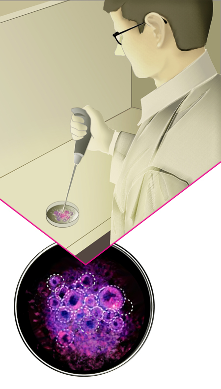
|
Illustration: Jane Kim |
By placing the iPS cells in different media, he nurtures the cells through their transformation. It is a process he has learned as much through trial and error as by design. When small tubes begin to form in the Petri dish, Pasca knows he is on his way. These are “rosettes,” which under the microscope look like luminescent clusters of corals. Their appearance marks the first visible stage of neuronal development. It’s enough to break Pasca’s calm demeanor.
“It is very exciting,” Pasca says. “I come in and say, ‘They should be forming rosettes now. They should be forming rosettes now. Will I have more tomorrow? And more, and more, and more?’”
Dolmetsch watches the developments with a keen eye. “All the results, even small results, he immediately pays attention to,” Pasca says. “’What can we do about it? What is it? It is valid? Is it not? What does it mean?’ Even for small, small things.”
Once the neural tube has formed, Pasca takes out the neural progenitors—the precursors to the brain cells—and triggers their transformation into neurons and glia. After several weeks, the neurons start to form connections. Finally, small bursts of electricity dart between them. They begin to talk. “If you had just asked me, 'Can we make a brain in a dish?,' I’d say ‘no’,” Dolmetsch says. “But in fact we can make little bits of brain in a dish in a really quite remarkable way.”
Eventually the cells die. It is impossible to keep them alive in the dish for more than a couple of months, Dolmetsch says. But they live long enough for the next stage of the experiment, Dolmetsch’s area of expertise. Dan Geschwind, a neurobiologist at UCLA's medical school, admires the work from afar. “It’s not just 'gee whiz, I can make these cells,'” Geschwind says. “The beautiful thing about Ricardo’s work is that he has these beautiful tools for looking at the cell biology and the physiology as well. He is on the forefront for sure.”
Cell talk
At the center of Dolmetsch's work is calcium, the wholesome ingredient of milk and strong bones. In the brain, calcium converts electricity into long-term changes in the structure and function of neurons. When an electrical signal reaches a neuron, channels in the cell membranes open and calcium ions flood into the cell. In Dolmetsch’s lab his students study the calcium signaling, or the opening and closing of pores, to try to detect any abnormalities. “It turned out that there are these mutations in calcium channels that are associated with a devastating form of autism,” Dolmetsch says.
Once his delicate brain tissues are ready, Pasca loads them with a calcium dye and observes them under a high-speed microscope. The microscope records and measures the rapid movement of calcium moving in and out of the cells. Seen on a computer screen, the results are mesmerizing. Small blotches of the green dye turn into tendrils reaching between the neurons. Suddenly the whole screen is a mass of green, only to recede just as quickly into quiet morass of black. This is the brain at work—or not.
Dolmetsch believes his research could be the first step in developing a drug or treatment for autism. Doctors prescribe drugs to treat individual symptoms, such as anxiety or aggression, but as yet no drugs exist to cure the disorder. “The standard line is you might be able to improve their symptoms, but they won’t get cured from their autism,” Dolmetsch says. “They are still autistic.”
The goal of his research is to identify drug targets: key molecules in the complex sequence of events of calcium signaling that a drug could influence. Snyder, however, offers words of caution. “One of the problems with the iPS field is that it forces you to become overly reductionist,” he says. “You are just working with a nerve cells in a dish and not a true patient.”
“That’s absolutely true,” Dolmetsch says. “But you have to be reductionist because there is no other way of studying brain diseases.” Since Dolmetsch began his autism research, several other groups, including Snyder’s, have also begun to use iPS cells to study the disorder.
Dolmetsch has already identified one possible drug target and believes that more will follow. “It is somehow starting to seem more tractable,” he says.
When he first embarked on the research, the challenge seemed such an “enormous mishmash of different things,” and he had his doubts. Now, he believes that for children with a specific defect in one class of cells, the future is hopeful. “I do believe there will be treatments,” he says. However, he admits his research is not a panacea. Many autistic children’s symptoms cannot be tied to known genetic mutations.
As for his own child? Dolmetsch is still for several seconds and then replies quietly. “I don’t know that I will be able to treat my son,” he says.
Reference
1. Leo Kanner. "Autistic Disturbances of Affective Contact," Nervous Child 2 (1943): 217-250. Reprinted in Childhood Psychosis: Initial Studies and New Insights.
Story ©2010 by Jane Palmer. For reproduction requests, contact the Science Communication Program office.
Top
Biographies
 Jane Palmer Jane Palmer
B.S. (cognitive science) University of Sheffield, U.K.
Ph.D. (computational molecular biology) University of Sheffield, U.K.
Internship: Cooperative Institute for Research in Environmental Sciences, University of Colorado, Boulder
My Indiana Jones tendencies bewitched me into science. Entranced by the physical world, I wanted to dig beneath the surface and uncover its endless mysteries. In academia, I burrowed into computer science, psychology, biology, and environmental science; outside, I rock-climbed across the continents. Eventually, a more traditional career path ensnared me, but the narrow focus and mundanities of research stifled my free spirit. And then, science writing liberated me.
When I write about science, I still get to hypothesize and explore. But it can be about Saturn one day, stem cell research the next, and water pollution the day after that. Better yet, as a writer, I polish my treasures for all to see.
With words as my tools, the adventure is only just beginning.
. . . . . . . . . . . . . . . . . . . . . . . . . . . . . . . . . . . . . . . . . . . . . . . . . . .
 Eva Langhorst Eva Langhorst
B.A. (fine arts) Gerrit Rietveld Academie, Amsterdam, Netherlands
I have always liked to work in great detail. When I was a child I made little models of our house. I liked to create anything that was small and precise. Later, during my study at the Art Academy in Amsterdam, both art and science often inspired my work. For my thesis project I worked in collaboration with a biologist to create small glass balls in which we planted enclosed biospheres. But my real fascination for science illustration came later, when my father, a dentist, asked me if I wanted to make a textbook about periodontology with him. The project ended up lasting years, but it was a great way to learn how art and science can come together. The experience led me to pursue science illustration as a career.
. . . . . . . . . . . . . . . . . . . . . . . . . . . . . . . . . . . . . . . . . . . . . . . . . . .
 Jane Kim Jane Kim
B.F.A. (printmaking) Rhode Island School of Design
Like all artists of science, the natural world has always inspired and excited me. As a kid, I survived a number of small obsessions including drawing flowers, raising pets—turtles, fish, birds, cats, dogs—sculpting horses, making and collecting teddy bears, insects in jars, and drawing portraits of dogs. Art was a clear choice. My post-college journey has been filled with many rich and fulfilling artistic endeavors, from working as printer at Paulson Press to carrying out an artist-in-residence at Recology in San Francisco. Reflecting on these different paths, I see that my work is bound together by a common thread—inspiration sparked by nature. I am driven by my vision to conserve and protect the natural world where I feel most excited, both visually and intellectually. Combining that with my passion for art leaves me with unrefined excitement.
For the next nine months, I look forward to many exciting science art adventures. My journey begins in Yosemite National Park through which I was awarded the Sierra Nevada Research Institute Science Visualization Fellowship. Following this experience, I am honored to work with The Smithsonian Tropical Research Institute in Panama. I will be working at Coiba National Park and the Naos Marine Lab. Finally, I will travel to Cornell University’s Lab of Ornithology where I was awarded the Bartels Science Illustration Internship.
Please visit me at www.ink-dwell.com and browse through Renderings of Life—a glimpse into places in which I like to dwell
Top |

